Scan-free stimulated Raman scattering tomography for label-free volumetric and functional deeper tissue and cells imaging
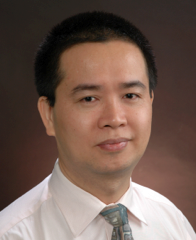
主讲人: Zhiwei Huang, PhD
时 间: 5月17日上午10:30
地 点: 光电大楼光子芯片研究院1131会议室
Abstract
Stimulated Raman Scattering (SRS) microscopy has become an appealing imaging tool for label-free biomolecular imaging in biological and biomedical systems. The conventional SRS microscopy uses the tightly focused Gaussian beams to excite the biosamples, which suffer from the strong light scattering in turbid media (e.g., tissue), resulting in a very limited penetration depth unsuited for acquiring deep tissue information. In this talk, I will present a novel z-scan-free SRS tomography (SRST) technique based on the optical beating method associated with non-diffracting Bessel beams to mitigate the scattering effect, enabling deeper tissue 3D imaging. The working principle of SRST technique and the method for retrieving the depth-resolved 3D information in frequency-domain will be explained and discussed. Further, I will also show some recent results of SRST technique for structural and functional 3D imaging in living tissue and cells. We demonstrate that the z-scan-free optical sectioning ability of optical beating technique developed in SRST is universal, which can be readily extended to any other imaging modalities for advancing 3D tissue and cells imaging in biomedical systems.
Brief Biography

Dr Zhiwei Huang is Associate Professor with tenure in the Department of Biomedical Engineering (BME), College of Design and Engineering at the National University of Singapore (NUS). He is a world-renowned expert in biomedical optics, biophotonics and microscopy imaging. His major research areas are in the fields of biomedical optics, microscopy, Raman spectroscopy and imaging, particularly centering on the development of super-resolution microscopy and nonlinear optical microscopy imaging techniques (e.g., coherent Raman scattering microscopy, multiphoton microscopy) and their applications in biomolecular imaging, as well as the development of novel fiber-optic Raman spectroscopy and endoscopic imaging, enabling early diagnosis and detection of epithelial precancer and cancer at endoscopy. He has pioneered in Raman endoscopy and label-free super-resolution bioimaging technologies, and published over 120 peer-reviewed articles in leading journals (e.g., Nature Photonics, Gastroenterology), and delivered over 100 plenary/keynote/invited lectures worldwide. He has filed over 20 US patents with 10 licensed for commercialization. He won many international awards related to novel Raman spectroscopy and imaging techniques. His IMDX technique invented was ranked No.1 among the top 10 medical devices listed in Medica, Germany. He is Elective Fellow of SPIE-the international society for optics and photonics.


